Saturday, June 29, 2013

Pasta

A rare state of matter dubbed "nuclear pasta" appears to exist only inside ultra-dense objects called neutron stars, astronomers say.
There, the nuclei of atoms get crammed together so tightly that they arrange themselves in patterns akin to pasta shapes — some in flat sheets like lasagna and others in spirals like fusilli. And these formations are likely responsible for limiting the maximum rotation speed of these stars, according to a new study.
"Such conditions are only reached in neutron stars, the most dense objects in the universe besides black holes," said astronomer José Pons of Alicante University in Spain. 
This new phase of matter had been proposed by theorists years ago, but was never experimentally verified. Now, Pons and his colleagues have used the spin rates of a class of neutron stars called pulsars to offer the first evidence that nuclear pasta exists.
Pulsars emit light in a pair of beams that shoot out like rays from a lighthouse. As the pulsars spin, the beams rotate in and out of view, making the stars appear to "pulse" on and off, and allowing astronomers to calculate how fast the stars are spinning.
Researchers have observed dozens of pulsars, but have never discovered one with a spin period longer than 12 seconds. "In principle, that is not expected. You should see some with larger periods," Pons told SPACE.com. A longer spin period would mean the star is spinning more slowly.

But the pasta matter could explain the absence of pulsars with longer spin periods. The researchers realized that if atomic nuclei inside the stars were reorganizing into pasta formations, this matter would increase the electric resistivity of the stars, making it harder for electrons to travel through the material. This, in turn, would cause the stars' magnetic fields to dissipate much faster than expected. Normally, pulsars slow their spin down by radiating electromagnetic waves, which causes the stars to lose angular momentum. But if the stars' magnetic fields are already limited, as would happen with pasta-matter, they cannot radiate electromagnetic waves as strongly, so they cannot spin down.

Whale

The blue whale is not only the largest animal alive currently, but is likely the largest animal ever to have lived.


Depending on the location, blue whales can grow to be 33 meters (108 feet) and weigh as much as 181 metric tons (200 imperial tons).They can live to be 90 years old and ingest about 3.6 metric tons (4 imperial tons) of krill each day. Blue whale are considered endangered by the IUCN.

Friday, June 28, 2013

Lego

Last year, two Canadian boys sent a LEGO Minifigure up to the stratosphere on a homemade rig. For only $400, they were able to connect a weather balloon to a platform that held four digital cameras, a parachute, and a LEGO man, proudly holding the Canadian flag. This wasn’t done for a school project; the boys did it in their spare time purely to see if they could. Their project was a success, and the apparatus went up an estimated 24 kilometers in the air; triple the height of a commercial airliner.

University of Toronto astrophysics professor Dr. Michael Reid hailed the boys’s experiment as extremely resourceful and likened it to work that Ph.D. students perform.


Thursday, June 27, 2013

Bacteria


Ground-breaking research by Australian scientists has revealed new insights into how life-threatening bacteria colonise medical devices that are implanted in the human body.
The break throughs could help tackle antibiotic resistant infections that develop in groups of microorganisms where cells stick to each other on a surface, otherwise known as biofilms. 
Biofilms are a significant threat to human and animal health because they are notoriously resistant to antibiotic treatment and immune defences. They are responsible for many chronic and recurring infections that occur around implanted medical devices.
Research published today in the prestigious journal Proceedings of the National Academy of Sciences USA (PNAS) by UTS Associate Professor Cynthia Whitchurch, used advanced microscopy techniques, including the next generation imaging system OMX Blaze, and sophisticated computer vision analysis to explore how bacterial biofilms spread. Associate Professor Whitchurch is leading a team of researchers from the UTS ithree institute, the UTS School of Mathematical Sciences, Monash University, University of Melbourne and CSIRO.
Studying the bacteria Pseudomonas aeruginosa, a pathogen that can cause respiratory disease and often causes infections when things such as catheters are inserted, has helped the team deduce how individual cell movements are co-ordinated and how intricate networks of interconnected trails are formed during biofilm expansion.
“This research reveals how co-ordination between individual bacteria can lead to complex group behaviour to enable biofilm expansion on a surface” says Associate Professor Whitchurch.
The researchers discovered that groups of bacteria build and migrate along a “furrow” on a surface – much like a well-trodden path across a grassy field. The team also discovered that extracellular DNA (eDNA) acted not as coding molecules but as a structural ‘rope’ that helped guide the transit of the bacteria.
What they discovered, in effect, was a traffic management system similar to signalling on a railway track that ensured a smooth flow of bacterial cells down the line towards the front of an expanding biofilm. eDNA also acted as glue, binding groups of bacteria together and forming ‘bulldozers’ that forged the furrows. 
Through its ithree institute, UTS has invested in world leading imaging facilities alongside state-of-the-art genomic, bioinformatics and proteomic facilities. 
The ground-breaking research demonstrated how such investment in world class facilities in NSW was advancing science as well as paving the way for its application to new medical discoveries, said Director of the ithree institute Professor Ian Charles.
"With the rise of resistant superbugs, we desperately need to find new ways to combat infectious diseases and I am confident that our research will make a contribution to that urgent quest" said Professor Charles.
The ithree institute is pioneering the use of OMX 3D SIM super resolution microscopy to advance the understanding of infectious disease. It is the world’s first commercial site for the next generation DeltaVision OMX Blaze super resolution imaging system – a device developed and manufactured in the United States by Applied Precision, a GE Healthcare Company. It is capable of capturing real-time multiple colour images of interactions between microorganisms and living cells.

Wednesday, June 26, 2013

Squid


Luckily, it has razor sharp teeth to properly chew its meals.

It also has the largest eyeball in the animal kingdom, measuring up to 25 cm (10 inches).

Sugar cube


The number plate is the chemical equation for Glucose, and the car is a white Cube. Someone had fun with this.

Theory


Definition of a scientific theory: a well-substantiated explanation of some aspect of the natural world, based on a body of knowledge that has been repeatedly confirmed through observation and experiment.

Other modern scientific theories: germ theory, theory of relativity, heliocentrism, atomic theory, plate tectonics, and cell theory.

PS Yes, gravity is a theory. Look up "the theory of gravity" and "Newton's Law Of Gravitation". Also, a theory never becomes a law.

Monday, June 24, 2013

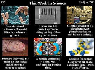
This week in science


Device that detects disease


An NJIT research professor known for his cutting-edge work with carbon nanotubes is overseeing the manufacture of a prototype lab-on-a-chip that would someday enable a physician to detect disease or virus from just one drop of liquid, including blood. "Scalable nano-bioprobes with sub-cellular resolution for cell detection," , (Elsevier, Vol. 45), which will publish on July 15, 2013 but is available now online, describes how NJIT research professors Reginald Farrow and Alokik Kanwal, his former postdoctoral fellow, and their team have created a carbon nanotube-based device to noninvasively and quickly detect mobile single cells with the potential to maintain a high degree of spatial resolution.


"Using sensors, we created a device that will allow medical personnel to put a tiny drop of liquid on the active area of the device and measure the cells' electrical properties," said Farrow, the recipient of NJIT's highest research honor, the NJIT Board of Overseers Excellence in Research Prize and Medal. "Although we are not the only people by any means doing this kind of work, what we think is unique is how we measure the electrical properties or patterns of cells and how those properties differ between cell types."

Saturday, June 22, 2013

New species


Typhochlaena costae.

A new brazilian tarantula


Much species exploration is undertaken out of pure curiosity about the living world, so it is easy to understand why progress in some obscure taxa has been slow. Other groups, however, are so significant that neglect of their taxonomy is inexplicable. Theraphosidae is one such taxon.
The theraphosids include about one-third of the 2,693 documented species of mygalomorphs – tarantulas and funnel web spiders. In the intricate sub-webs of invertebrate animals in many terrestrial ecosystems these are "top" predators, making them of special interest to ecology. They have been used as model organisms in such diverse studies as molecular biology, biochemistry, pharmacology, ethology and biogeography, and they have a large and devoted following in the international pet trade. Yet their taxonomy, natural history and phylogeny remain incompletely studied.
Professor Rogério Bertani, of the Instituto Butantan, São Paulo, recently revised three theraphosid genera, improving our knowledge of their species diversity, phylogeny, and geographic and natural histories, and describing nine species as new. Although there were more interesting natural history stories attached to some of his other new species, I could not get past the sheer beauty of one, Typhochlaena costae. Its spectacular colouration, long golden setae (hairs) over the chelicerae (jaws), carapace and abdomen are chelicerae-dropping gorgeous.


Silver

Though silver has been used to help treat wounds for years, new research shows that when added to antibiotics, it can be 10-1,000 times more effective than using the antibiotic alone. Researchers hope that this could be used to help attack the ever-growing list of antibiotic-resistant bacteria. Silver helps compromise the membrane of bacteria, allowing the medication to be more effective.


The antimicrobial properties of silver have been known for thousands of years, but it was not clear how the metal wreaked havoc on pathogenic invaders. Now, researchers have explained the cellular processes by which the precious metal weakens bacteria and makes them more susceptible to antibiotics, according to a study published on June 19 in Science translational magazine. The findings suggest that silver could be used to enhance the effectiveness of antibiotics against drug-resistant bacteria.
A team lead by Jim Collins, a biomedical engineer at Boston University, showed that dissolved silver ions interfere with several cellular processes in bacteria, including disulfide-bond formation, iron homeostasis, and metabolism. These changes not only make the cell membrane more permeable, but also lead to increased production of reactive oxygen species, which can induce cell death via DNA damage. 

Solar Energy

In January and February alone, the United States produced 730 million kilowatt hours of electricity from solar panels. This is an astonishing 10% of all of the solar electricity generated during 2012. Considering those are two of the least sunny months of the year, it sets expectations for summer months very high.


3D brain

The model increases resolution by a factor of 50 when compared to images provided by an MRI. Individual layers of the brain model had to be stacked and lined up perfectly. It took 4 years to align the 7,404 slices that were only 20 µm thick. Altogether, the scientists gained a terabyte of data on the brain.

rock


Friday, June 21, 2013

Particle accelerator

The accelerator was developed at the University of Texas and is scaled down by a factor of 10,000 when compared to conventional accelerators. Within a decade, an accelerator of 20 gigaelectronvolts may be possible. This design has the potential to revolutionize research by making technology available to institutions that do not have access to traditional units due to space or budget constraints


"We have accelerated about half a billion electrons to 2 gigaelectronvolts over a distance of about 1 inch," said Mike Downer, professor of physics in the College of Natural Sciences. "Until now that degree of energy and focus has required a conventional accelerator that stretches more than the length of two football fields. It's a downsizing of a factor of approximately 10,000."


Downer said he expects 10 GeV accelerators of a few inches in length to be developed within the next few years, and he believes 20 GeV accelerators of similar size could be developed within a decade.
Downer said that the electrons from the current 2 GeV accelerator can be converted into "hard" X-rays as bright as those from large-scale facilities. He believes that with further refinement they could even drive an X-ray free electron laser, the brightest X-ray source currently available to science.
A tabletop X-ray laser would be transformative for chemists and biologists, who could use the bright X-rays to study the molecular basis of matter and life with atomic precision, and femtosecond time resolution, without traveling to a large national facility.
"The X-rays we'll be able to produce are of femtosecond duration, which is the time scale on which molecules vibrate and the fastest chemical reactions take place," said Downer. "They will have the energy and brightness to enable us to see, for example, the atomic structure of single protein molecules in a living sample."

More info: http://bit.ly/16k1S8F

For Science!!!!


I am going to share the latest revelations in science on this blog.
-Arjun